About one in eight women in the U.S. will develop invasive breast cancer during her lifetime, according to the American Cancer Society. Fortunately, it is no longer the probable death sentence it once was, in large part because today’s well-designed screening tests help doctors find breast tumors at an earlier stage, thus increasing the chances for successful treatment.
Prof. Degani is developing a new method, DTI, for early detection of breast cancer and, potentially, pancreatic cancer. DTI is completely non-invasive, as it tracks water in tissue to detect cancer.
“If you detect breast cancer very early, you make it a disease that is in more cases curable. And that’s really our aim,” says Prof. Hadassa Degani of the Weizmann Institute of Science’s Department of Biological Regulation. For more than 20 years, she has been developing less-invasive cancer diagnosis techniques that utilize existing magnetic resonance imaging (MRI) scanners.
One of these methods, known as 3TP (Three Time Point), received FDA approval in 2003 for use in the detection of breast and prostate cancers. It enables doctors to distinguish between benign and malignant tumors at very early stages, sometimes even when tumors can’t be detected by other methods.
The technique consists of injecting a contrast-enhancing dye-like material that circulates in the patient’s bloodstream, then using MRI to produce high-resolution images of the tumor at three judiciously preselected times: before the contrast material is injected and twice afterwards, at intervals of several minutes. The way in which the dye-like material is taken up and cleared out by the tumor tissue is monitored in the scanner; this, in turn, reveals the amount of blood microvessels and their permeability, as well as the cells’ density, making it possible to distinguish between malignant and benign tumors, and to determine the potential aggressiveness of a malignant tumor.

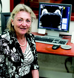
Prof. Hadassa Degani
Unlike mammography scanners, MRI scanners don’t expose patients to harmful X-ray radiation. MRI-based screening techniques also have the potential to reduce the number of biopsies performed, sparing patients pain and anxiety and lowering costs.
To adapt the 3TP technique to diagnose prostate cancer, Prof. Degani and her team had to determine the optimal time points for obtaining the three MRI images of the prostate gland. Until recently, the only way to definitively diagnose prostate cancer was to do biopsies on tissue taken from up to eight different sites.
A significant number of scanners sold today to hospitals and clinics in the U.S. and worldwide, including Israel, enable the use of the 3TP method for diagnosing breast or prostate cancer, under license from iCAD Ltd. But Prof. Degani says that she hopes that MRI-based cancer screening techniques will be even more widely used in the future, so that more patients can benefit from the high accuracy of the 3TP method.
While many hospitals and clinics today use 3TP to diagnose breast and prostate cancer, Prof. Degani hopes its usage will spread further, giving even more patients the benefit of the technique’s high accuracy.
Now she is developing another promising diagnostic method – one that is completely non-invasive, as it requires no injections of contrast material. Known as diffusion tensor imaging (DTI), the approach involves tracking the natural movement of water molecules in tissues in all directions. Using DTI, Prof. Degani and her team survey the network of mammary ducts in the breast.
“Breast cancer always starts in the ducts and glandular elements. They get blocked by cancer cells,” explains Prof. Degani. “With DTI, we can see the ‘ductal trees’ and identify their blockage by cells through changes in the water’s movement.”
A major advantage of the DTI method is that it can be used in some groups of women for whom mammography or the 3TP technique would either be unsafe or ineffective – for example, women who are pregnant, lactating, treated with hormone replacement therapy, or suffering from renal failure.
“We realized that this approach can be very powerful, and patented it,” notes Prof. Degani. She is now working to apply the DTI method to detect pancreatic cancer, which is often only discovered in its later stages.
“With the support of donors, we were able to build a whole new facility for MRI research. Most of my research today is done on people, which is new for me.”
When Prof. Degani first developed her 3TP technique, she worked with collaborators in the U.S. who conducted the clinical trials. Now, after conducting initial development and clinical studies of the DTI method on the Weizmann campus, she is moving to conduct clinical trials in centers in Israel and abroad. “With the support of donors, we were able to build a whole new building for MRI research. Most of my research today is done on people, which is new for me,” she says.
Prof. Degani received her MSc in physical chemistry from the Weizmann Institute’s Feinberg Graduate School and her PhD in chemistry from the State University of New York at Stony Brook. She joined the Weizmann faculty in 1976 and began studying the role of blood vessel formation in tumor growth and metastasis – in particular, how blood vessel formation is regulated by hormones. The 3TP method arose from those early investigations.
“I started with very basic research – exploring questions that had nothing to do with the end point,” she says. “But asking these questions helped me find answers that I hope will have a real impact on cancer.”
